The procedure: dual-plane endoscopic augmentation
Source: Partner clinic technical brief (breast augmentation materials, received 2026-08-02)
What is dual-plane augmentation?
In a dual-plane augmentation the implant sits partly behind the pectoralis (chest) muscle and partly behind the breast gland, creating two layers of support — combining secure implant coverage with natural movement, while lowering the probability of capsular contracture.
What each plane does
- Plane one (behind the gland): the upper part of the implant sits against the breast tissue — natural shape, soft feel;
- Plane two (behind the muscle): the lower part is covered by muscle — less visible implant edge, lower risk of sagging or displacement;
- The key manoeuvre: releasing the lower attachment of the pectoral muscle with precisely controlled scope — over-dissection would let the implant ride up.
What do the endoscope and ultrasonic scalpel add?
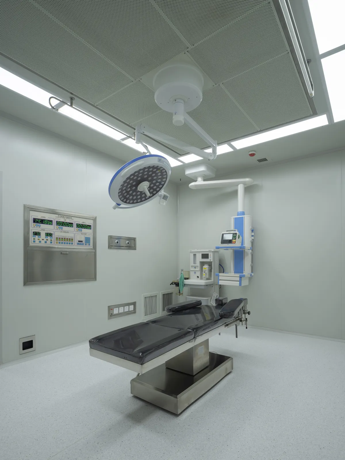
The partner clinic operates fully visualized through an endoscope, using an ultrasonic scalpel instead of conventional electrocautery for muscle release, plane dissection and haemostasis:
- Dissection under direct vision — avoids the vessel and nerve injuries of blind dissection; the two implant pockets come out more symmetrical;
- The ultrasonic scalpel cuts and seals simultaneously — intraoperative bleeding is far lower than in conventional augmentation and the swelling phase is shorter;
- Better protection of the intercostal nerves and chest-wall vessels — markedly lower rates of post-operative numbness;
- Precise pocket dissection — significantly fewer long-term complications such as capsular contracture and implant displacement.
The three incision routes
| Incision | Character | Suits |
|---|---|---|
| Armpit (transaxillary) | Most concealed; endoscope-friendly view | Scar-averse patients with a reasonable breast base |
| Lower areola edge | Short dissection path; most precise dual-plane control | Mild sagging, thinner gland tissue |
| Breast fold (inframammary) | Most direct, plane in full view; visible scar | Less commonly chosen in China |
The route is decided with your surgeon at the consultation, based on your anatomy and priorities.
What actually happens during surgery
- Access: a small armpit or areola incision; the endoscope establishes a visualized working channel;
- Layered dissection: the ultrasonic scalpel first opens the retropectoral space and releases the muscle fibres precisely, then the retroglandular space; the dual-plane boundary is kept near the nipple line;
- Haemostasis and pocket refinement: cutting and sealing happen together so the field stays dry; both pockets are matched for size and depth;
- Implant placement: sterile insertion, orientation and base position adjusted, symmetry confirmed, pocket thoroughly irrigated to minimize foreign-body reaction;
- Drain and closure: a suction drain is placed, tissue closed in layers, compression dressing fixes the implant position.
Safety lines the surgical team holds
- No forceful blunt dissection — protects against muscle tearing and implant displacement;
- The ultrasonic scalpel never dwells on one spot — prevents heat injury that could later trigger contracture;
- The sensory nerves of the nipple-areola are protected throughout to avoid permanent numbness.
The information on this site is for general education only, drawn from materials supplied by our partner clinic. It is not medical advice, does not create a doctor–patient relationship, and cannot tell you whether any procedure is right for you. Only a licensed physician who has examined you can do that.
Ask a coordinator about your case